Invited state-of-the-art review
Knee dislocations with vascular injuries – a review article
26. feb. 2024
9 min.
Abstract
Key Points
“Of this I have only seen one instance, and I conclude it, therefore, to be a rare occurrence; and there are scarcely any accidents to which the body is liable which more imperiously demand immediate amputation than these” [1]. This is a statement on knee dislocation by Sir Astley Cooper from 1824, who is usually credited to be the first to report on these injuries. Similar sentiments were echoed by William Gibson in 1825 when he wrote: "Complete luxation of the knee was extremely rare and was generally followed by violent symptoms and even death unless treated by timely amputation." To some this might sound dramatic, but it highlights the seriousness of the injury and its consequences if not treated in a timely manner. Some of the reasons why amputation was the treatment of choice may include vascular injuries, open injuries with contamination and, of course, lack of aseptic surgical techniques at the time (1824). Despite advances in modern technology and surgical techniques, late diagnosis and treatment of vascular injuries will usually result in amputation, which can be lifesaving but devastating for the patient. Therefore, early diagnosis and treatment of vascular injuries is paramount. Interestingly, a recent study from the Mayo Clinic emphasised the seriousness of vascular injuries as they reported that patients with knee dislocation and concomitant vascular injuries had poorer outcomes than those without vascular injuries [2].
Knee dislocation injuries were historically believed to be a relatively rare, reported to constitute 0.02-0.2% of orthopaedic injuries. The true incidence of knee dislocations remains unknown but is probably higher because the diagnosis can be challenging, and some knee dislocations may be misdiagnosed because about 50% are reported to reduce before presentation [3, 4]. Interestingly, the risk of neurovascular injuries is similar in patients presenting with dislocated knees and those presenting with reduced bicruciate ligament injured knees [3, 4]. This highlights the need for high level of suspicion and meticulous and systematic evaluation of these patients.
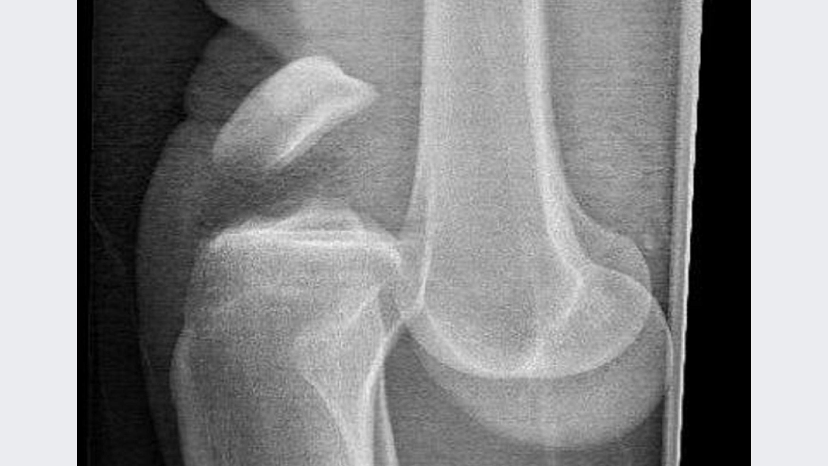
Historically, knee dislocations have been associated with high-energy trauma such as motor vehicle accidents, falls from heights and farm/industry injuries, resulting in frank knee dislocation (Figure 1). In the 19th and early 20th century, the mechanisms of injury usually included a cart and horse falling on its owner, a man on horseback whose leg was pinned between a rail and the horse from which he was being thrown and, less often, some less dramatic injury mechanisms such as falls. With the advent of motorised vehicles, industrialisation and sports participation, the mechanisms of injury have changed. In recent years, increasing attention has been devoted to knee dislocations in morbidly obese patients, often termed ultra-low velocity knee dislocations. These patients often sustain a knee dislocation during daily activities or a fall from their own height. The morbidly obese patients also represent a special treatment group because they have a higher risk of concomitant neurologic and vascular injuries [5].
Knee dislocations are complex injuries and are often associated with other injuries. The rate of associated injuries to other vital organs such as the head, chest and/or abdomen has been reported to be 27% in high-energy trauma. Fractures are also commonly associated with knee dislocations, reported in the 50-60% range; and the rate of multiple fractures has been reported to be 41%. The prevalence of concomitant fractures in the ipsilateral limb is high (17-58%) and may influence the decision making in the initial treatment of these injuries. Patients with bilateral multi-ligamentous knee injuries are reported to have a higher risk of concomitant head, chest and abdominal injuries than unilateral multi-ligamentous knee injuries with similar mechanisms.
The rate of popliteal artery injury has been reported to fall in the 7-48% range, and to be as high as 64% in cases of fractures, depending on the individual hospital reporting [6]. Level 1 trauma centres, where the majority of patients with knee dislocations have high-energy trauma, usually report a higher prevalence of concomitant vascular injuries. The risk of vascular injury is correlated with the degree of energy, concomitant fractures and the type of dislocation. Peroneal nerve injury is associated with vascular injury. Therefore, the presence of peroneal nerve injuries should be considered indicators for potential vascular injuries. Merritt et al. reported that 38% of patients presenting with common peroneal nerve palsy had a concomitant arterial injury, and 62% of the patients with knee instabilities associated with arterial injuries had transient or permanent peroneal nerve neuropraxia. This implies that vascular injuries should be suspected in patients with nerve injuries because of the associated risk and the difficulty in assessing vascular perfusion in patients with nerve injuries. The threshold for vascular assessment with angiography should be low because the consequences of missing a vascular injury may be devastating for the patient. Data from Oslo University Hospital showed that 5-6% of patients with knee dislocations sustain a concomitant vascular injury. Interestingly, the odds for popliteal artery injury were nine times higher among those with knee dislocations involving the lateral structures than among those with other ligament injury types. Additionally, peroneal nerve injury was significantly associated with vascular injury (odds ratio = 20.6; 95% confidence interval: 5.3-118.8) [7].
It is paramount to know the anatomy of the knee to understand the risk for vascular structures around the knee. The popliteal artery (Figure 2) courses through the adductor hiatus, where there are fibrous attachments to the tendinous attachment of the adductor magnus muscle, “securing” the artery in place. The popliteal artery has branches which form the superior genicular, middle genicular and the inferior genicular arteries. In the popliteal space, the artery has relatively little structural attachment, making this section relatively mobile. It is the deepest or most anterior structure in the popliteal fossa, separated from the knee joint capsule by a thin layer of fat tissue (Figure 2 and Figure 3). When it passes distally, the artery is anchored to the proximal tibia by the fibrous arch of the soleus muscle. Thus, the artery is tethered at this point before it divides into the anterior and posterior tibial arteries. Because of the firm attachments proximal and distal to the popliteal space, when the knee dislocates, the portion of the artery in the popliteal space is susceptible to injury. Contusion injuries to the artery in this section may also occur in case of a direct trauma. Injuries may also occur if there is a concomitant fracture because of the close proximity to both the femur and tibia in the popliteal space.
Pulses are routinely utilised to assess vascularity and the presence of vascular injuries. The presence of pulses in patients without neurologic injuries and hypovolaemia can be used to exclude vascular injuries. However, it should be noted that the presence of pulses does not exclude vascular injuries. The examination of pulses alone has been demonstrated to be unreliable in detecting vascular injuries, leading some authors to recommend routine angiography for a knee dislocation. In patients with abnormal or asymmetric pulses, an ankle-brachial index (ABI) measurement has been recommended (Figure 4). The utility of measuring ABI has some limitations, including cases of knee dislocation associated with ipsilateral limb fracture or hypovolaemic shock. Furthermore, an ABI cannot detect vascular intimal lesions that may lead to a secondary occlusion even if the ABI initially appears to be normal. An ABI < 0.9 warrants further examination with angiography [8] because ABI < 0.9 had a 95-100% sensitivity and a 80-100% specificity in detecting vascular lesions requiring surgery. Standard angiography has 1.2-6% false negatives, up to 7% false positives and is associated with complications. However, the risk of complications with angiography seem to outweigh the potential risk of limb loss when the diagnosis of arterial injury is delayed or missed. With recent developments in imaging, CT angiography is preferred to standard angiography owing to low complication rates and high sensitivity [9] (Figure 5).
Vascular injuries require urgent treatment; and, when suspected, a vascular surgeon should be contacted. Further evaluation after performing ABI should be discussed with the vascular surgeon. However, further investigations in a hospital without vascular surgery care should not delay transfer and treatment.
Treatment of vascular injuries usually involve repair of the popliteal artery utilising a venous graft. Fasciotomy is performed to mitigate development of a reperfusion compartment syndrome (Figure 6). A spanning external fixator is used to stabilise the limb during surgery and is maintained for 4-6 weeks after surgery to stabilise the knee and protect the repair. After 4-6 weeks, the spanning external fixator is removed and a brace is used to stabilise the knee. Then the external fixator is removed, manipulation under anaesthesia might be indicated if there is severe stiffness. In cases of vascular injuries requiring repair, ligament reconstruction is delayed to approximately six months.
Concomitant vascular injuries during knee dislocations are not uncommon and require timely diagnosis and treatment. Delay exceeding eight hours may lead to amputation, which is physically and emotionally devastating for the patient. ABI should be performed in all patients with knee dislocations, and a low threshold for CT angiography to avoid delay in treatment. Vascular surgeons should be contacted early when vascular injury is suspected because vascular repair takes precedence over ligament repair.
Correspondence Lars Engebretsen. E-mail: lars.engebretsen@medisin.uio.no
Accepted 10 January 2024
Conflicts of interest Potential conflicts of interest have been declared. Disclosure forms provided by the authors are available with the article at ugeskriftet.dk/dmj
Acknowledgements The authors take this opportunity to express their gratitude to Kirurgen.no for permitting this manuscript to be published in the Danish Medical Journal. Previously published 5 January 2023 in Kirurgen.no.
Cite this as Dan Med J 2024;71(3):A08230529.
doi 10.61409/A08230529
Open Access under Creative Commons License CC BY-NC-ND 4.0